Difference Between Lung Infiltrate and Consolidation
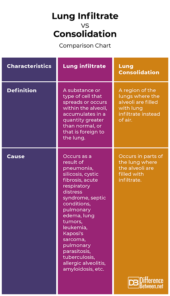
When we breathe in, the inhaled air enters the trachea, the branches, and through progressively smaller airways reaches the alveoli – microscopic bubbles, where the gas exchange occurs. In some cases, the alveoli can fill up with a substance, for example with pus (e.g. in pneumonia), protein (in certain rare lung diseases), blood, water (e.g. pulmonary edema), cancer cells, etc. This substance is called lung infiltrate. The alveoli filled with infiltrate can be seen on a chest X-ray as an area, denser than the surrounding lung tissues, called consolidation.
What is Lung Infiltrate?
Any substance or type of cell that spreads or occurs within the alveoli, accumulates in a quantity greater than normal, or is foreign to the lung is called lung infiltrate. The infiltrate can be composed of white blood cells, cancer cells, protein, pus, blood, etc.
Lung infiltration can occur in a large number of diseases, including pneumonia, asbestosis, silicosis, cystic fibrosis, actinomycosis, acute respiratory distress syndrome, septic conditions, pulmonary edema, lung tumors, leukemia, Kaposi’s sarcoma, pulmonary parasitosis, tuberculosis, nocardiosis, staphylococci, rheumatoid arthritis, dermatomyositis and polymyositis, scleroderma, sarcoidosis of the lungs, allergic alveolitis, amyloidosis, etc.
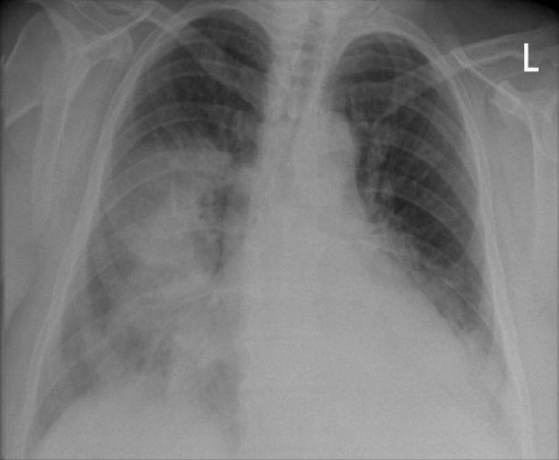
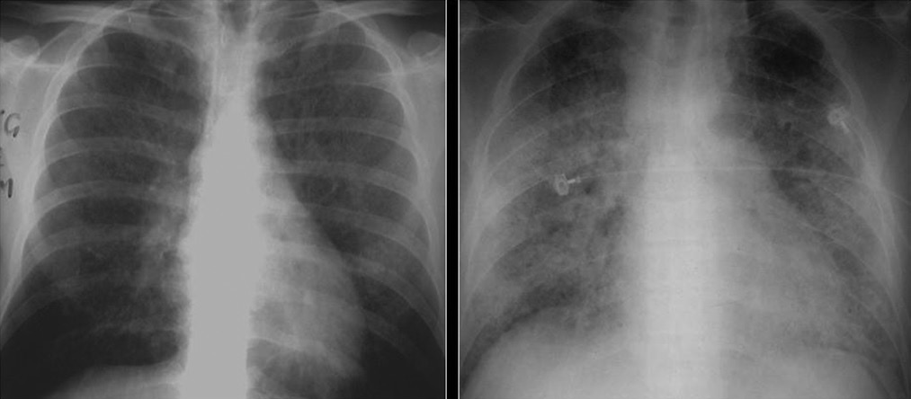
What is Lung Consolidation?
The region of the lungs where the alveoli are filled with an atypical substance (i.e. lung infiltrate) instead of air is called lung consolidation. It is marked by hardening or swelling of the lung tissue and obstruction of the aeration in the affected area of the lung. Consolidation occurs in different diseases (when lung infiltrate occurs) and is an important radiologic sign.
Depending on the severity of the condition, the consolidation can cover a very small area of the lung surface and cause minimal symptoms, or cover a large part of the lung and cause a life-threatening condition.
Radiographs of the lungs and symptoms are similar in consolidation resulting from all types of infiltrate. Therefore, more research is usually needed to determine the exact cause of the condition. Consolidation of the lungs makes it difficult to breathe. Air cannot pass through the small airways, so the lungs cannot function properly. Symptoms, depending on the cause of the condition, may include:
- Shortness of breath;
- Cyanosis;
- Thick green or bloody sputum;
- Cough with blood;
- Dry cough;
- Noisy breathing;
- Chest pain or heaviness;
- Rapid breathing;
- Fever;
- Fatigue, etc.
Some of the common causes of consolidation are pneumonia, pulmonary edema, and pulmonary hemorrhage.
Pneumonia is the most common cause of lung consolidation. When there is an infection in the lungs, the body sends white blood cells to fight it. Dead cells and debris accumulate, creating pus that fills the small airways. Pneumonia is usually caused by bacteria or viruses, but can also be caused by fungi or other causes.
Congestive heart failure is the most common cause of pulmonary edema. When the heart cannot pump enough to keep the blood moving, it returns to the blood vessels in the lungs. The increased pressure pushes fluid out of the blood vessels into the small airways.
Pulmonary hemorrhage is most often caused by vasculitis (inflammation of the blood vessels), which makes the blood vessels weak and causes blood to pass into the small airways.
With proper treatment, pulmonary consolidation usually disappears and air is returned to the small airways.
Difference Between Lung Infiltrate and Consolidation
Definition
Lung infiltrate: Any substance or type of cell that spreads or occurs within the alveoli, accumulates in a quantity greater than normal, or that is foreign to the lung is called lung infiltrate.
Lung Consolidation: The region of the lungs where the alveoli are filled with an atypical substance (i.e. lung infiltrate) instead of air is called lung consolidation.
Cause
Lung infiltrate: Lung infiltration can occur in different diseases, including pneumonia, silicosis, cystic fibrosis, acute respiratory distress syndrome, septic conditions, pulmonary edema, lung tumors, leukemia, Kaposi’s sarcoma, pulmonary parasitosis, tuberculosis, allergic alveolitis, amyloidosis, etc.
Lung Consolidation: Lung consolidation occurs in parts of the lung where the alveoli are filled with infiltrate.
Lung Infiltrate Vs Consolidation: Comparison Chart
Summary:
- Any substance or type of cell that spreads or occurs within the alveoli, accumulates in a quantity greater than normal, or that is foreign to the lung, is called lung infiltrate.
- The region of the lungs where the alveoli are filled with lung infiltrate instead of air is called lung consolidation.
- Lung infiltration can occur in different diseases, including pneumonia, silicosis, cystic fibrosis, acute respiratory distress syndrome, septic conditions, pulmonary edema, lung tumors, leukemia, Kaposi’s sarcoma, pulmonary parasitosis, tuberculosis, allergic alveolitis, amyloidosis, etc.
- Lung consolidation occurs in parts of the lung where the alveoli are filled with infiltrate.
- Depending on the severity of the condition, the consolidation can cover a very small area of the lung surface and cause minimal symptoms, or cover a large part of the lung and cause a life-threatening condition.
- With proper treatment, pulmonary consolidation usually disappears and air is returned to the small airways.
- Difference Between Gallstones and Cholecystitis - September 5, 2021
- Difference Between Constipation and Cramping - August 4, 2021
- Difference Between Whole Genome Sequencing and Microarray - May 6, 2021
Search DifferenceBetween.net :
Leave a Response
References :
[0]Des Jardins, T., G. Burton. Clinical Manifestations and Assessment of Respiratory Disease, 7th Edition. St. Louis, Missouri. 2016. Print.
[1]Warrell, D., T. Cox, J. Firth. Oxford Textbook of Medicine, Vol. 2. Oxford: Oxford University Press. 2010. Print.
[2]Yankova, Z. New Guide to Pulmonary Diseases and Tuberculosis. Sofia: Medical University Press. 2012. Print.
[3]Image credit: https://commons.wikimedia.org/wiki/File:X-ray_lung_consolidation.jpg
[4]Image credit: https://commons.wikimedia.org/wiki/File:X-ray_of_ground_glass_opacities_of_pneumocystis_pneumonia.jpg