Difference between ECG and Echocardiography
Introduction:
Electrocardiogram (EKG or ECG) and Echocardiography are painless, non-invasive tests used to evaluate the functioning of the heart. These tests are usually ordered by the physician, performed by a technician or the physician himself after which the test result is interpreted. Both these tests do not require prior preparation and do not carry any risks for the patient.
Difference in technique:
ECG is a recording of the electrical activity of the heart. This is done by attaching painless electrodes that can record this activity to the surface of the skin. 12 patches are stuck to the chest, arms and legs which are connected by wires to a machine. This machine displays the electrical activity on a paper for interpretation. The procedure takes not more than 10 minutes and does not involve any electric shocks, or harm to the body. An ECG can also be conducted during exercise to look for result of stress on the heart.
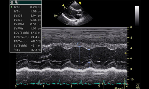
Echocardiography is a test that uses sound waves to create images of the beating heart. It uses the standard 2-dimensional, 3-dimensional, and Doppler ultrasound. Echocardiography may be performed transthoracic (from over the chest), transesophageal (by introducing a recorder in the food pipe) or as stress echocardiography. The doctor performs the test by moving a device called a transducer over the chest which is connected to a monitor which captures images of the heart. The procedure takes not more than 10-15 minutes.
Difference in uses:
An ECG records the electrical activity of the heart and thus gives valuable information about the rate at which the heart beats, and the rhythm and regularity of the heartbeat. An ECG is a quick screening method done to detect arrhythmias, damage to the heart muscle during a heart attack, condition of any device implanted like a pacemaker and diagnosis of certain congenital conditions and effects of drugs. An ECG is also used as a routine health check up and also is a part of the work up done before any major surgery.
Echocardiography provides a wide range of information of the heart regarding its size, shape, the pumping capacity, location and extent of tissue damage, internal chambers of the heart, functioning of the valves. It is used to determine the condition of the heart muscle after a heart attack mostly. It can detect infection of the sac around the heart and infection over the valves of the heart. A color Doppler echocardiogram can give an accurate assessment of the blood flowing through the heart.
Summary:
Electrocardiogram and Echocardiography are extremely helpful tests used to diagnose several conditions of the heart. ECG records the electrical activity of the heart whereas echocardiography uses sound waves to create pictures of the heart. An ECG can detect irregularities in the pace and rhythm of the heartbeats. Echocardiography gives much more added and detailed information about the structure as well as functioning of the heart muscle and its valves. An ECG takes hardly 10 minutes whereas echocardiography is a slightly lengthy procedure depending on the type of condition of the heart. However, both these tests are extremely safe and easy to perform.
- Difference between near sightedness and far sightedness - January 21, 2015
- Difference between Diverticulosis and Diverticulitis - January 20, 2015
- Difference between Prilosec and Nexium - January 19, 2015
Search DifferenceBetween.net :
Leave a Response
References :
[0]http://upload.wikimedia.org/wikipedia/commons/b/b3/PLAX_Mmode.jpg