Difference Between Macule and Papule
What is Macule and Papule?
Both are skin conditions. Both are dermatological manifestations (skin problems) which occur in various local or systemic (something that is spread throughout) disease conditions.
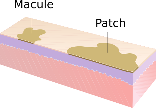
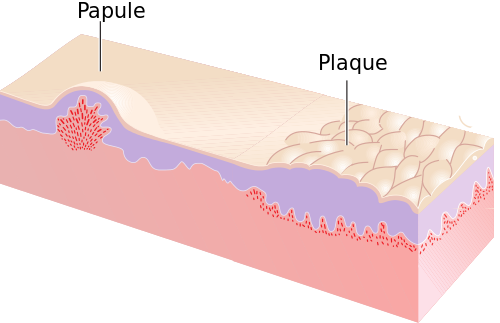
However, macule is a non-elevated discolored skin patch on the skin and papule is an elevated white lesion that is smaller than 0.5cm in diameter.

What is Macule?
A small (usually < 1 cm in diameter) flat, non-palpable and altered in color – a discolored blemish or lesion that can be brown, tan, red or white and has same texture as surrounding skin.
Macular rashes usually happen in following medical conditions
- Reactions due to some medications
- Inflammatory conditions like allergies
- Infections by viruses such as EBV (Epstein-Barr virus) and bacteria such as hepatitis

What is Papule?
It is an elevated, circumscribed solid cutaneous lesion that is up to 1 cm in size. It can be present in variety of colors like red brown, purple brown, brown, pink, and yellow brown.
It can occur on the face, often on the eyelids or alar rim, but also on the trunk and extremities.
Difference between Macule and Papule
Definition
Macule
A macule is a discolored patch of skin, flat, distinct, neither raised (elevated) nor depressed – less than 1 centimeter (cm) wide.
Macule doesn’t alter the texture and thickness of the skin. These blemishes or skin discoloration is caused by various causes. For example, macules could be vitiligo lesions (can be lighter or hypopigmented) or moles (that are darker in color or hyperpigmented).
Papule
Papules are cutaneous, circumscribed, inflamed blemishes sometimes and solid elevation of the skin, up to 1 cm in diameter. Papules are tiny lesions or dome-shaped elevations on the skin that are either red or pink colored and can be well seen on the skin. Papules have well-defined boundaries and usually occur on the face. Shapes of papules could be different. Sometimes these papules (elevated bumps on the skin) cluster together to form what appears like a rash and feels rough like sandpaper. Papules often start as erythematous macules.
Causes
Macule
Macules can be caused by various conditions that impact the way your skin color and texture look, leading to discolored areas. Conditions that are likely to cause macules are:
- Vitiligo (patches of skin losing their pigment)
- Moles (common non-cancerous skin growth usually dark brown triggered by clusters of pigmented cells)
- Freckles (tiny brown patches on your skin, usually in areas that get exposed to the sun)
- Sun spots, age spots, and liver spots
- Post-inflammatory hyperpigmentation (PIH) – dark spots left behind after a pimple heals such as that which appears after acne lesions heal
- Tinea versicolor (a common fungal infection of the skin)
Papule
The primary causes of papules (raised bumps or elevations on skin) in general, include:
- Too much oil production
- Bacterial infection
- Increased activity of male sexual hormones (androgens)
- Leishmaniasis
- Different forms of vasculitis
Acne (Papules) can also be triggered or aggravated by:
- Anxiety and stress
- Processed foods or diet, (consumption of too much sugar)
- Certain drugs like corticosteroids
Size
Macule
Flat papule <1 cm
Papule
Raised macule up to 1 cm
Etiology and layers of skin involved
Macule
Only dermis
Papule
Proliferation of cells in epidermis or superficial cells
Type of lesion
Macule
Flat area of altered color or texture
Papule
Elevated solid lesion
Examples
Macule
Nevi, actinic keratoses, sun spots, age spots, warts, lichen planus, freckle, insect bites, liver spots, seborrheic keratoses, some lesions of acne, tinea versicolor, and skin cancers.
Papule
Warts, lichen planus, rash, insect bites, seborrheic keratoses, acne pimples or lesions, actinic keratoses, warts, bumps (white blood cells attacking an infection) and skin cancers.
Summary
The points of difference between Macule and Papule have been summarized as below:

- Difference Between Global Warming and Greenhouse Effect - May 18, 2024
- Difference Between Vaccination and Immunization - March 3, 2024
- Difference Between Selective Mutism and Autism - February 25, 2024
Search DifferenceBetween.net :
Leave a Response
References :
[0]Aldahan, A. S., Brah, T. K., & Nouri, K. (2018). Diagnosis and management of pearly penile papules. American journal of men's health, 12(3), 624-627.
[1]Fadok, V. A. (1995). Overview of equine papular and nodular dermatoses. Veterinary Clinics of North America: Equine Practice, 11(1), 61-74.
[2]Hori, Y., Kawashima, M., Oohara, K., & Kukita, A. (1984). Acquired, bilateral nevus of Ota-like macules. Journal of the American Academy of Dermatology, 10(6), 961-964.
[3]Lallas, A., Argenziano, G., Moscarella, E., Longo, C., Simonetti, V., & Zalaudek, I. (2014). Diagnosis and management of facial pigmented macules. Clinics in dermatology, 32(1), 94-100.
[4]Image credit: https://upload.wikimedia.org/wikipedia/commons/thumb/a/ab/Macule_and_Patch.svg/500px-Macule_and_Patch.svg.png
[5]Image credit: https://upload.wikimedia.org/wikipedia/commons/thumb/1/1e/Papule_and_Plaque.svg/500px-Papule_and_Plaque.svg.png
