Difference Between Somatic and Autonomic Nervous System
Introduction
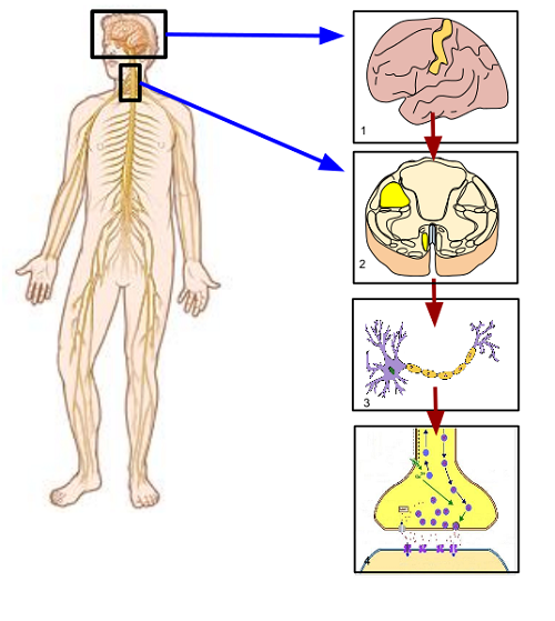
The peripheral nervous system is an extension of the central nervous system. Its overall function is to carry information from the central nervous system to other parts of the body to maintain normal body function. It enables the body to react voluntarily and involuntarily to any stimuli. It is composed of nerve fibers bundles that lie beyond the brain and spinal cord. Some of the nerve fiber bundles proceed to innervate skeletal muscles and sensory receptors. These fibers comprise the somatic nervous system. The remaining nerve fibers innervate visceral organs, smooth muscles, glands and blood vessels. These fibers comprise the autonomic nervous system.
Somatic Nervous System
The somatic nervous system is composed of nerves that originate from the spinal cord. Nerves that supply muscles on the head originate from the brain. It is comprised of motor neurons that supply skeletal muscles to allow movement. Its axon is continuous from the spinal cord to the skeletal muscle, forming the neuromuscular junction. The neuromuscular junction is an important structure for neurotransmission to stimulate muscular contraction. Inhibition of locomotion occurs through inhibitory pathways coming from the central nervous system.
Transmitters and Receptors
The space between the motor neuron and the skeletal muscle is called a synaptic cleft. The axon terminal of motor neurons releases the neurotransmitter, acetylcholine, which is the only neurotransmitter for the somatic nervous system. Acetylcholine is stored within vesicles located on the knob-like terminal end of the nerve fiber, called a terminal button. The terminal button contains calcium channels. When calcium is sufficiently released, this triggers the release of acetylcholine from the vesicles into the synaptic cleft. Acetylcholine binds to nicotinic cholinergic receptors, which activates a series of chemical reactions that changes the ionic composition of the motor endplate.
Effector Organs and Function
The release of acetylcholine stimulates the opening of ionic channels for sodium and potassium. Ionic particles carry an electrical charge and concentration gradient. This reaction generally moves sodium inward and potassium outward causing a depolarization of the motor end plate. This allows electrical current to flow from the depolarized motor end plate and adjacent areas triggering the opening of voltage-gated sodium channels. This propagates an action potential throughout the effector organ, which is the skeletal muscle. The initiated electrical potential activity spreads within the entire muscle allowing contraction of the skeletal muscle fiber. The aforementioned chain of events enables voluntary control of muscle groups that is essential for locomotion.
Autonomic Nervous System
The autonomic nervous is system is composed of nerves that originate from the brain and the spinal cord. It is also known as the visceral nervous system because its nerve bundles proceed to supply visceral organs and other internal structures. Its axon is discontinuous and is separated by a ganglion, forming a two-neuron chain. The autonomic nervous system has two functionally different subdivisions. The sympathetic division enables the human body to involuntary respond to emergency situations, creating a “fight or flight” response. The parasympathetic division enables normal visceral functions by allowing storage of energy to conserve body reserves.
Transmitters and Receptors
The autonomic nervous system preganglionic neurons release acetylcholine at the synaptic area, which binds to nicotinic cholinergic receptors at the postsynaptic membrane. In parasympathetic nervous system, post-ganglionic neurons also release acetylcholine, which binds to muscarinic receptors located in salivary glands, stomach, heart, smooth muscles and other glandular structures. In sympathetic nervous system, post-ganglionic neurons release norepinephrine, which binds to alpha-1 receptors in smooth muscles, beta-1 receptors in the heart muscle, beta-2 in smooth muscles and alpha-2 adrenergic receptors.
Effector Organs and Function
Both the sympathetic and parasympathetic nerve fibers are present in all visceral organs. The principal effector organs that regulate homeostatic organs are the skin, liver, pancreas, lungs, heart, blood vessels and kidneys. Nerve fibers from the sympathetic and parasympathetic subdivisions are complementary in function to allow involuntary mechanisms that preserve the internal homeostatic mechanisms. The skin serves to regulate the body’s core temperature by preserving or conserving water loss from sweat glands. The liver and the pancreas regulate the metabolism of glucose and lipids. The lungs regulate the concentration of oxygen and acidic particles in the blood by allowing oxygen inhalation and carbon dioxide exhalation. The heart and blood vessels regulate blood pressure through cardiac rhythmic nodes and blood vessel wall diameter changes. The kidneys regulate the excretion of toxins in the body. It also works synergistically with the lungs to maintain normal blood pH levels.
Summary
The somatic and autonomic nervous systems have salient anatomic and structural differences that give rise to different functions. Somatic nerves predominantly come from the spinal cord and are composed of motor neurons that travel to the skeletal muscle. It releases acetylcholine, which stimulates the voluntary contraction of skeletal muscles. Its function is controlled by central nervous system structures such as the motor cortex, basal ganglia, cerebellum, brainstem and the spinal cord. On the other hand, autonomic nerves come from both the spinal cord and the brain that travels to various internal organs, smooth muscles, glands and blood vessels. It is comprised of a two-neuron chain with a preganglionic area that releases acetylcholine, and a post-ganglionic area that releases acetylcholine for parasympathetic terminals and norepinephrine for sympathetic terminals. Neurotransmitter release allows involuntary control of visceral organs by stimulation or inhibition. This is regulated by central nervous system structures such as the prefrontal cortex, hypothalamus, medulla and spinal cord.
- The Difference Between Imitrex and Relpax - February 18, 2017
- Difference Between Reactants and Products - November 15, 2016
- Difference Between Plants and Humans - October 13, 2016
Search DifferenceBetween.net :
1 Comment
Leave a Response
References :
[0]Berne, R. M., Koeppen, B. M., & Stanton, B. A. (2010). Berne & Levy physiology. Philadelphia, PA: Mosby/Elsevier.
[1]Preston, R. R., & Wilson, T. E. (2013). Physiology. Philadelphia: Wolters Kluwer Health/Lippincott Williams & Wilkins.
[2]Sherwood, L. (2012). Fundamentals of Human Physiology. Belmont, CA: Brooks/Cole Cengage Learning.
[3]Snell, R. S. (2010). Clinical Neuroanatomy. Philadelphia: Wolters Kluwer Health/Lippincott Williams & Wilkins.
[4]Weiten, W. (2012). Psychology: Themes and Variations. Belmont, CA: Cengage/Wadsworth.
[5]https://en.wikipedia.org/wiki/Somatic_nervous_system
Nice website, love it.