Difference Between Apical and Lateral Meristematic Tissue
According to their main purpose, plant tissues are divided into:
- Meristematic (forming) tissues – ensure the growth of plants;
- Permanent tissues – perform all other functions.
Meristematic cells divide, forming new cells that increase in size and differentiate, forming all plant tissues and organs.
According to their origin, meristematic tissues are divided into primary and secondary.
- Primary meristematic tissues – originate from the seed germ (apical meristems, pericycle);
- Secondary meristematic tissues – originate later in the life of the plant, most often from dedifferentiated parenchyma cells, which regain the ability to divide (cambium, phylogeny).
According to their location, meristematic tissues are divided into:
- Apical – located at the tips of the root, their branches. Cause the increase of the roots, stems, and branches in length;
- Lateral – located laterally in the axial organs of the plant. Provide thickening of the root and stem in shrubs, trees, and some herbaceous plants;
- Intercalary – inserted among more or less differentiated tissues. Provide elongation of the plant organs.

What is Apical Meristematic Tissue?
Apical meristematic tissue is a meristematic tissue located at the top of the root, stem, and their branches, causing the growth of plant organs in length. It originates from the meristem cells of the embryo and is primary in origin. Apical meristematic tissue is found in all higher plants.
The cells of the apical meristematic tissue are not equivalent. The initial cell or group of initial cells, together with the adjacent derivative cells, is referred to as the protomeristem. The cells of the protomeristem have the ability to divide an unlimited number of times and retain their meristematic character until the end of the plant’s life. However, cell division is not limited to the area of the apical meristematic tissue. It also occurs in more distant and partially differentiated meristematic tissues, known as primary (transitional) meristems. These transitional meristems – protoderm, main meristem, and procambia – are formed by the activity of the apical meristematic tissue and form cylindrical layers. The cells of the transitional meristems remain meristematic for some time, after which, mainly according to their location, they differentiate into cells of the three tissue systems – ground, vascular, and dermal.
Depending on its location, the apical meristematic tissue is:
- Root apical meristematic tissue;
- Stem apical meristematic tissue.
The root apical meristematic tissue forms new cells and contributes to the growth of roots and their branches in length, providing plants with access to water and nutrients.
The stem apical meristematic tissue is located in the apical and lateral buds of the stem and twigs. In addition to forming new cells and increasing the length of the stem and twigs, it also forms:
- Leaf primordia –which develop into leaves;
- Primordia of lateral buds – which develop into lateral branches.

What is Lateral Meristematic Tissue?
The lateral meristematic tissue is a meristematic tissue located on the lateral side of the stems and roots, causing the growth of plant organs in thickness. They can be of primary or secondary origin.
The lateral meristematic tissue occurs only in trees, shrubs, and some grasses.
The lateral meristematic tissues are:
- Cambium;
- Phellogen (cork cambium);
- Procambium;
- Pericycle.
The lateral meristematic tissue forms cylinders located parallel to the long axis of the stems and roots.
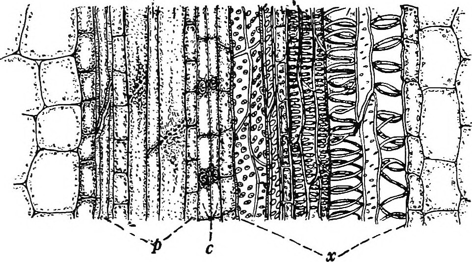
The cambium forms the secondary conducting tissues – secondary phloem and secondary xylem. It is a cylinder of several layers of cells – one layer of initial cells and their closest derivatives.
It consists of two types of cells:
- Spindle-shaped initial sells – elongated cells, pointed at their ends;
- Ray initial sells – almost isodiametric small cells.
The cells of the cambium divide tangentially. The cells produced outwards from the cambial ring differentiate into cells of the secondary xylem, and those separated inwards – into cells of the secondary phloem.
Cambium cells are secondary in origin and are derived from procambia cells in the conducting bundles and dedifferentiated parenchymal cells located between the conducting bundles.
The phellogen (cork cambium) forms the secondary covering tissue – cork. It is laid the primary bark of the root and stem. The first embedded phylogeny is always a cylinder of several layers of thin-walled cells with a rectangular shape. Its cells divide tangentially and form numerous layers of cortical cells on the outside and several layers of phelloderm cells on the inside. It is with a secondary origin and is derived from dedifferentiated cells of the epidermis or parenchyma of the primary cortex.
The pericycle forms a cylinder of one or more layers of meristem cells, the first layer of the central cylinder of the root. It forms the lateral branches of the root.
Difference Between Apical Meristematic Tissue and Lateral Meristematic Tissue
Definition
Apical meristematic tissue: Apical meristematic tissue is a meristematic tissue located at the top of the root, stem, and their branches, causing the growth of plant organs in length.
Lateral meristematic tissue: The lateral meristematic tissue is a meristematic tissue located on the lateral side of the stems and roots, causing the growth of plant organs in thickness.
Origin
Apical meristematic tissue: Apical meristematic tissues are of primary origin.
Lateral meristematic tissue: Lateral meristematic tissues are of primary or secondary origin.
Occurrence in plants
Apical meristematic tissue: Apical meristematic tissue is found in all higher plants.
Lateral meristematic tissue: Lateral meristematic tissue occurs only in trees, shrubs, and some grasses.
Location
Apical meristematic tissue: Apicalmeristematic tissue is located at the top of the root and stem and their branches.
Lateral meristematic tissue: The lateral meristematic tissue forms cylinders located parallel to the long axis of the stems and roots.
Types
Apical meristematic tissue: Depending on its location, the apical meristematic tissue is root apical meristematic tissue or stem apical meristematic tissue.
Lateral meristematic tissue: The lateral meristematic tissues are cambium, phellogen (cork cambium), procambium, pericycle.
Growth
Apical meristematic tissue: The apical meristematic tissue contributes to the growth of roots, stems, and their branches in length, forms primordia of leaves and lateral buds.
Lateral meristematic tissue: The lateral meristematic tissue is responsible for the plant widening/thickening.
Formed tissues
Apical meristematic tissue: The apical meristematic tissue forms the xylem, phloem, epidermis, and ground tissue.
Lateral meristematic tissue: The lateral meristematic tissue forms wood, inner bark, and outer bark.
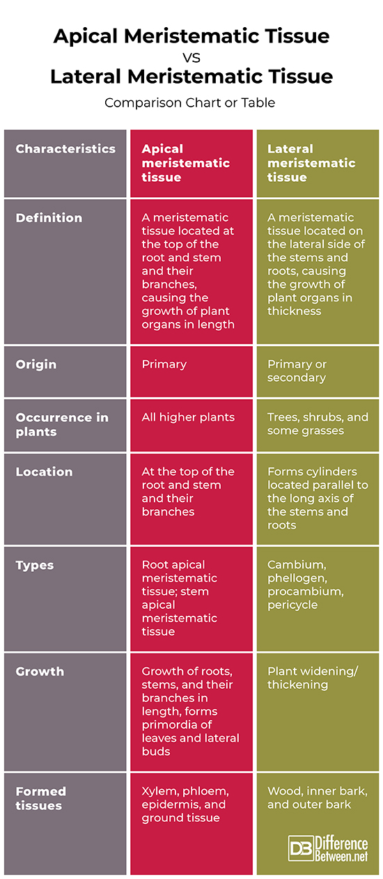
Apical Meristematic Tissue Vs. Lateral Meristematic Tissue: Comparison chart or table

Summary:
- According to their main purpose, plant tissues are divided into meristematic tissues, which ensure the growth of plants and permanent tissues, which perform all other functions.
- According to their location, meristematic tissues are divided into apical, lateral, and intercalary.
- Apical meristematic tissue is a meristematic tissue located at the top of the root and stem and their branches, causing the growth of plant organs in length.
- The lateral meristematic tissue is a meristematic tissue located on the lateral side of the stems and roots, causing the growth of plant organs in thickness.
- Apical meristematic tissues are of primary origin. Lateral meristematic tissues are of primary (procambium, pericycle) or secondary origin (cambium, phellogen).
- Apical meristematic tissue is found in all higher plants. Lateral meristematic tissue occurs only in trees, shrubs, and some grasses.
- Apical meristematic tissue is located at the top of the root, stem, and their branches. The lateral meristematic tissue forms cylinders located parallel to the long axis of the stems and roots.
- The apical meristematic tissue is root apical meristematic tissue or stem apical meristematic tissue. The lateral meristematic tissues are cambium, phellogen (cork cambium), procambium, pericycle.
- The apical meristematic tissue contributes to the growth of roots, stems, and their branches in length, forms primordia of leaves and lateral buds. The lateral meristematic tissue is responsible for the plant widening/thickening).
- The apical meristematic tissue forms the xylem, phloem, epidermis, and ground tissue. The lateral meristematic tissue forms to wood, inner bark, and outer bark.
- Difference Between Gallstones and Cholecystitis - September 5, 2021
- Difference Between Constipation and Cramping - August 4, 2021
- Difference Between Whole Genome Sequencing and Microarray - May 6, 2021
Search DifferenceBetween.net :
Leave a Response
References :
[0]Barthelemy, D., Caraglio, Y. Plant architecture: a dynamic, multilevel and comprehensive approach to plant form, structure and ontogeny. London: Annals of Botany. 2007: 375–407. Print.
[1]Evert, Ray. Esau's Plant Anatomy: Meristems, Cells, and Tissues of the Plant Body: Their Structure, Function, and Development. Hoboken: John Wiley & Sons, Inc., 2006. Print.
[2]Neuhaus, G.. Morphology and Anatomy of Vascular Plants. Berlin: Springer. 2013. Print.
[3]Image credit: https://live.staticflickr.com/702/20778174742_fe4256dfec_b.jpg
[4]Image credit: https://en.wikipedia.org/wiki/Evolutionary_history_of_plants#/media/File:Angiosperm_life_cycle_diagram-en.svg
