Difference between Conjunctiva and Sclera
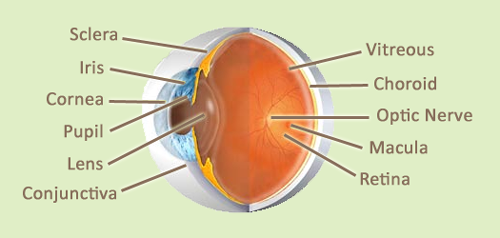
Eyes are one of the most vital sense organs of the human body as they are responsible for vision and nonverbal communication. The human eye is composed of a thick white layer called as the sclera and a thin translucent layer called as the conjunctiva. Both these layers are important to maintain the structure and function of the eye. Let us understand how both of these work to maintain normal sight.
The Conjunctiva:
The term conjunctiva gets its name from the word Conjoin which means to join. This is a thin, translucent membranous structure that covers the eye ball. It extends backwards to cover the sclera and folds upon itself and comes forwards by lining the under surface of the eyelid. At the margin of the eyelid it is continuous with the skin. The conjunctival fold allows unrestricted eyeball movement. The conjunctiva has its own nerve and blood supply. It is made up of non – keratinised stratified squamous epithelium and stratified columnar epithelium. The main function of the conjunctiva is to keep the eye lubricated by producing mucus and some amount of tears. It also prevents foreign particles and microbes from gaining entry into the eye. The contact lenses are held in place due to the presence of conjunctiva.
Parts of conjunctiva
The conjunctiva is divided into palpebral, bulbar and fornix conjunctiva.
The palpebral conjunctiva is located on the inside of the upper and lower eyelid.
The conjunctiva of the fornix is lining of the sac that is present at the junction between the posterior surface of the eyelid and the anterior part of the globe of the eye. The conjunctiva is relatively thicker and loose here which allows for free movement of the eyeball. The conjunctival sac formed at the transition of palpebral and fornix conjunctiva holds around 7 µl of tear fluids. The sac has a capacity to hold 30 µl of fluid.
The bulbar conjunctiva is the thinnest part of the conjunctiva. It covers the cornea and the anterior portion of the eyeball. It is so transparent that one can see the underlying white sclera and the blood vessels clearly with the naked eye.
Diseases of the conjunctiva
The conjunctiva can get irritated and inflamed on contact with dust particles or as part of autoimmune reaction. It is also susceptible to infection resulting in conjunctivitis or pink eye as it is commonly known. Conjunctiva is also vulnerable to age related changes and can get detached from the underlying sclera. Conjunctival tumours are also known to occur in some people.
Sclera of the eye
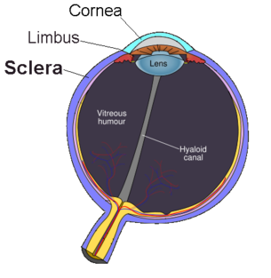
Sclera has its origins from the Greek word Skleros which means hard. The sclera is the tough outer coat of the eyeball. It is white in colour. The sclera is opaque as it is made up of white fibrous collagenous tissue. It is underlined by the choroid layer. The sclera is continuous with the cornea in the front and the optic sheath at the back of the eye. The optic sheath encircles the optic nerve as it emerges out from the retina. Inflammation of sclera is called as Scleritis which is a very rare phenomenon.
In humans the colour of the sclera contrasts with the smaller size and dark colour of the iris because of which it is distinctly visible. In other mammals the sclera gets camouflaged with the colour and larger size of the iris and is therefore not visible. The sclera is responsible for maintaining the shape of the globe and offers resistance to internal and external forces. It also provides a base for attachment for the extra ocular muscles that are responsible for eye movement. The thickness of the sclera varies from 1mm at the posterior most point to 0.3 mm just behind the rectus muscle insertions.
The sclera is also divided into four parts – episclera, stroma, lamina fusca and endothelium.
To summarise conjunctiva is a thin membrane that covers the front of the eye and the sclera is the thick white coat that forms the outer layer of the eyeball.

http://www.ericksonlabs.com/v/Artificial_Eyes/eye_conditions.asp
http://en.wikipedia.org/wiki/Sclera
- Difference Between Flu and Fever - September 2, 2015
- Difference Between Haemoglobin and Iron - July 10, 2015
- Difference between Purpura and Ecchymosis - July 2, 2015
Search DifferenceBetween.net :
4 Comments
Leave a Response
References :
[0]http://en.wikipedia.org/wiki/Conjunctiva
[1]http://en.wikipedia.org/wiki/Sclera
[2]http://www.ophthobook.com/chapters/anatomy
[3]http://www.eophtha.com/eophtha/Anatomy/anatomyofconjunctiva1.html
[4]http://www.ericksonlabs.com/v/Artificial_Eyes/eye_conditions.asp
Beautifully explained. Thanks.
This website completely confused me because it says the conjunctiva covers the cornea which is INCORRECT. When the lids are closed over the eye, maybe you could get away with saying this but it’s still misleading. The bulbar conjunctiva covers the sclera but stops at the cornea.
From a histology perspective, the cornea is simply a clear conjuctiva. Therefor the conjuctiva does not cover the cornea, but rather the cornea is a transparent continuation of the conjuctiva that was named seperately due to color.
You are right
That’s why in conjunctivitis redness stops at limbus