Difference Between the Morel-Lavallee Lesion and Hematoma
Morel-Lavallee lesions are soft-tissue degloving injuries secondary to trauma. These occur due to abrupt separation of epidermis and dermis from the imbedded fascia. Hematomas are soft, boggy swellings that occur due to pooling of blood in the injured tissue. Both these lesions are separate entities and should not be confused for one.
What is a Morel-Lavallee lesion?
Definition:
The Morel-Lavallee lesion first elaborated by a French surgeon Maurice Morel-Lavallee in 1853, is a post traumatic, closed degloving injury that occurs due to shearing force separating the dermis from the underlying fascia.
Causes:
High intensity forces to soft tissue are one of the most important causes of the Morel-Lavallee lesion. The underlying vasculature endures the injury and the inflammatory process begins in the tissue beneath. If left untreated in the acute stage, bacterial colonisation can take over leading to a complex lesion formation. Certain orthopedic conditions such as pelvic and acetabular fractures and proximal femur fractures are prone to developing the Morel-Lavallee lesion.
Diagnosis:
Physical examination is key to the diagnosis of Morel-Lavallee lesions. Radiological modalities are almost always used as these depict soft tissue swellings more evidently and detect any pelvic and acetabular fractures. CT scan of the area of interest is used to measure size of lesion. Ultrasound shows a hypoechoic space above the fascia and is a therapeutic modality if percutaneous treatment is needed. MRI is the most important test for evaluation of the Morel-Lavelle lesions.
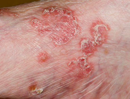
Symptoms:
These lesions are largely asymptomatic and up to 33% of lesions are missed on initial presentation. The patient may complain of a swelling of an area following trauma. Skin discoloration takes several days to become apparent. Fluctuance and paresthesia of overlying skin is noted on physical examination.
Treatment:
Non operative treatment options include compression therapy and percutaneous drainage with drain placement. These are limited to smaller lesions. Larger lesions require operative treatment. Single or dual incision irrigation and debridement is chosen from. Sometime chronic recurrent lesions are dealt with open debridement.
Complications:
Recurrence is the most common complication of Morel-Lavallee lesions. Chronic, untreated lesions can result in pseudocyst formation. Also, skin necrosis and perioperative infection are very frequent.
What is a hematoma?
Definition:
Hematoma is localized extravasation of blood from the capillaries which is secondary to a disease or trauma. Initially the blood persists in the liquid form in the tissue and later it coagulates and reabsorbs into the vessels.
Causes:
The primary cause of a hematoma is trauma. Other causes of hematoma include aneurysm, anticoagulants, viral infections, and orthopedic injuries such as fractures.
Diagnosis:
The diagnosis of hematoma primarily requires medical history and physical examination. Important tests that aid diagnosis include complete blood count, coagulation profile, metabolic panel, and liver function tests. Neurological hematomas are diagnosed via CT scan and MRI.
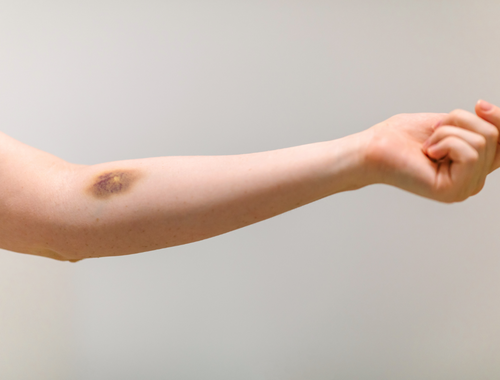
Symptoms:
The common symptoms of hematoma are pain, discoloration, swelling and erythema. Hematomas have different symptoms depending on their location. Neurological symptoms such as weakness on one side, difficulty in speaking, and confusion, are high alarm symptoms that need urgent evaluation as the hematoma might seeping through the brain tissue, like a time bomb. In subungual hematoma, nail pain, and nail disfiguration might be noticed.
Treatment:
Treatment options for hematoma may include rest, elevation of effected region, applying ice packs, and adequate pain relief. Rarely, surgical drainage may be needed in life threatening hematomas.
Complications:
Risk of infection is a common complication of hematoma.
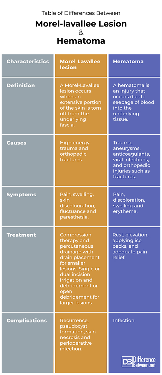
Difference between Morel-Lavallee lesion and hematoma
Definition:
A Morel-Lavallee lesion occurs when an extensive portion of the skin is torn off from the underlying fascia. A hematoma is an injury that occurs due to seepage of blood into the underlying tissue.
Causes:
High energy trauma and orthopedic fractures are common causes of Morel-Lavallee lesions. Hematoma can occur secondary to trauma, aneurysms, anticoagulants, viral infections, and orthopedic injuries such as fractures.
Symptoms:
Pain, swelling, skin discolouration, fluctuance and paresthesia are the symptoms of a Morel-Lavallee lesion. The common symptoms of hematoma are pain, discoloration, swelling and erythema.
Treatment:
Treatment options for Morel-Lavallee lesion include compression therapy and percutaneous drainage with drain placement for smaller lesions and single or dual incision irrigation and debridement or open debridement for larger lesions. Treatment options for hematoma include rest, elevation, applying ice packs, and adequate pain relief.
Complications:
Recurrence, pseudocyst formation, skin necrosis and perioperative infection are common complications of Morel-Lavallee lesions whereas infection is the main complication of hematoma.
Table of differences between Morel-Lavallee lesion and hematoma
FAQs:
Is Morel Lavallee lesion a hematoma?
No, a Morel Lavallee lesion is the separation of skin from underlying fascia whereas hematoma is an injury in which blood spills into the underlying tissue.
Where is the characteristic location of fluid as seen with a Morel-Lavallée lesion?
Between the subcutaneous fat and underlying fascia.
How do you get a Morel-Lavallée lesion?
A high impact, blunt force trauma can lead to the degloving lesion.
What is a morel Lavallee Seroma?
It is a complication of Morel-Lavallee lesion in which a pseudocyst forms.
How would you describe a degloving injury?
Separation of the skin from the fascia is a degloving injury.
How long does a Morel Lavallee lesion take to heal?
An average of 5 weeks.
Is a Morel Lavallee lesion painful?
Yes, it is a painful lesion.
Does a hematoma go away?
Most hematomas resolve quickly with conservative management. Some larger ones need surgical drainage.
What are 3 causes of a hematoma?
What does a hematoma look like?
A soft, boggy fluctuant swelling.
- Differences Between Reptiles and Amphibians - May 17, 2024
- Difference Between Ophthalmology and Optometry - May 15, 2024
- Difference Between Fear and Anxiety - April 2, 2024
Search DifferenceBetween.net :
Leave a Response
References :
[0]Nair, Anirudh V et al. “Morel-Lavallée lesion: A closed degloving injury that requires real attention.” The Indian journal of radiology & imaging vol. 24,3 (2014): 288-90.
[1]Myrick, Karen M., and Stephen Davis. "Morel‐Lavallee injury a case study." Clinical Case Reports 6.6 (2018): 1033.
[2]Eisele, David, and Richard V. Smith. Complications in Head and Neck Surgery E-Book. Elsevier Health Sciences, 2008.