Difference Between Necrosis and Eschar
Both necrosis and eschar indicate the unexpected death of cells when the supply of blood and oxygen is cut off to an organ or location of the body. Death of the cells is presumed to be linked to proteins inside the cell breaking down and the body releasing enzymes to digest the dead cells. Because the onset is unexpected, the body is not ready to remove the dead cells, so an inflammatory response ensues. If chemicals released affect adjacent cells, the necrosis spreads. Eschar, although also dead tissue, is a piece of hard, dark, dry tissue that has a leather-like and spongy appearance. Eschar cover the surface of the skin over a necrotic wound.
Causes of Necrosis
Necrosis has a wide range of causes. These include insufficient blood flow to bodily tissue or an organ, for example, due to deep vein thrombosis and physical injury; extreme temperatures that restrict blood flow, for example, frostbite; exposure to radiation in cancer treatments; contact with toxic chemicals or poisons, for example, spider and snake bites as well as some recreational drugs; and pathogenic micro-organisms like bacteria, fungi, and viruses that infect an organ, including the skin or bone.
Types of Necrosis
There are six types of necrosis according to Andréas Astier:
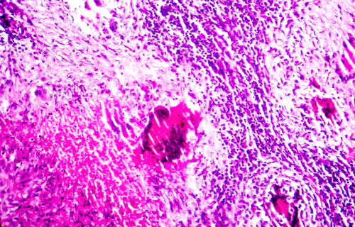
- Coagulative necrosis affects any tissue other than the brain and is caused by insufficient blood supply to the necrotic region.
- Liquefactive necrosis is the result of an enzyme imbalance that leads to cells digesting themselves. The wound appears pussy. Bacteria, fungi, viruses and parasites may cause this type of necrosis.
- Caseous necrosis is caused by the presence of noxious, foreign matter that is sealed off by the immune system to contain the damage.
- Fat necrosis occurs when pancreatic enzymes leak into and destroy the surrounding fatty tissue. The release of enzymes can be the result of viruses, infections, ischemia, trauma, and toxins.
- Fibranoid, or fibrin-like, necrosis occurs when nonglobular proteins responsible for blood clotting leak out of blood vessels.
- Gangrenous necrosis most often affects the extremities and may be dry when blood and oxygen fail to reach a region of the body or wet due to the presence of bacterial infection.
Treatments for Necrosis and Eschar
Necrosis and eschar can be treated with surgery aimed at removing the dead tissue, which is called debridement, and restoring blood flow to the region; antibiotics to treat or prevent infection; and wound treatment protocols. It is not possible to return necrotic tissue or eschar to health, however: The tissue is dead. If left untreated, the decomposing dead tissue and cell debris build up at or near the dead cells. The latter condition is called gangrene.
Difference between necrosis and eschar
Necrosis means that body tissue has died prematurely in an unregulated and unexpected fashion (SY Proskuryakov). The condition is most often detrimental and often proves fatal (DL Kasper). The appearance of eschar suggests the wound is at a more advanced stage, and while the eschar functions as a natural barrier to infection because it keeps bacteria from infecting a wound, it also prevents classification of the wound into stages as well as healing of the wound (Rachel Nall). If the eschar becomes unstable, in other words, becomes moist, oozes, and is loose and red, debridement or cutting the eschar away may be recommended.
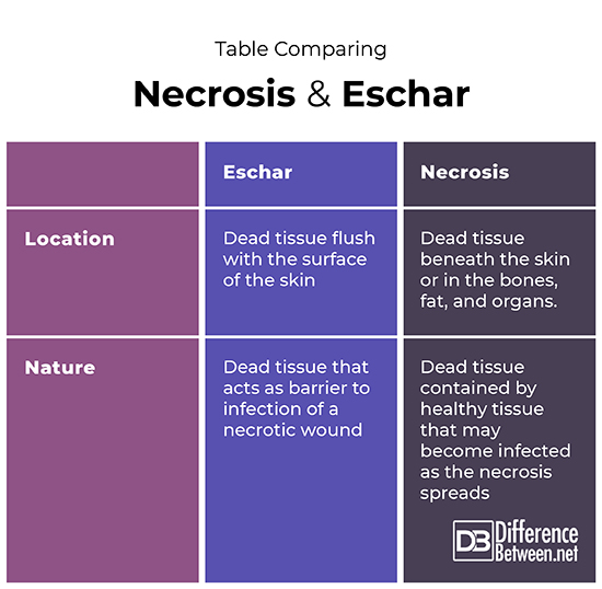
Table comparing necrosis and eschar
Summary
Necrosis is the result of blood and oxygen to affected tissue being obstructed and can occur in any organ of the body other than the brain, including the skin and bones. Eschar is a leathery and spongy covering that develops flush with the skin’s surface and covers the necrotic wound bed beneath. Its function is to protect the wound against infection.
FAQ
Is eschar and necrotic tissue the same thing?
Both eschar and necrotic tissue imply the unexpected and unregulated death of cells. While necrotic tissue can occur in any organ of the body, eschar appear flush with the skin surface and cover a wound bed of necrotic tissue. Eschar function to protect the underlying necrotic tissue from further infection by pathogens.
What does necrotic tissue eschar look like?
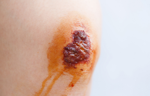
A stable eschar sits flush with the skin’s surface. It is a dry, leathery and spongy black, brown or tan tissue that covers necrotic tissue. If it becomes unstable, or soggy and red, it must be debrided or cut out (Cheryl Carver).
How can you tell the difference between a scab and eschar?
An eschar and scab are not the same. While both are often dry, eschar are necrotic or dead tissue whereas a scab is made up of dried blood and oozing serum. While a scab appears as a rusty brown crust on top of the skin surface that falls off naturally once the skin underneath has healed, a stable eschar is flush with the skin surface and consists of dead cells. Removal of an eschar risks exposing the underlying necrotic wound to infection by pathogens.
Is gangrene and eschar the same?
Gangrene is a coagulative necrosis that looks like tissue that has mummified and occurs when cells in a substantial area of a limb or parts of the gastrointestinal tract die because of insufficient oxygen and blood supply. If infection of the dead tissue occurs, it becomes liquefactive necrosis or wet gangrene. In contrast, eschar cover the underlying necrotic wound bed to protect the dead tissue from further infection by pathogens.
What does eschar look like?
An eschar is tan, brown, or black crusty tissue that resembles a piece of steel wool and appears flush with the skin. The eschar hides a necrotic would and the area surrounding the eschar may be inflamed and tender to the touch.
- Difference Between Ecchymosis and Erythema - August 15, 2022
- Difference Between Autobiographical Memory and Episodic Memory - August 1, 2022
- Difference Between Biological Drive and Social Motive - July 30, 2022
Search DifferenceBetween.net :
Leave a Response
References :
[0]Astier, Andréas. "The Different Types of Necrosis and Their Histological Identifications" [Blog]. Educating in Medicine & Pharmacy, June 30, 2020, https://www.andreasastier.com/blog/the-different-types-of-necrosis-and-their-histological-identifications.
[1]Carver, Cheryl. "Knowing the Difference Between Scabs and Eschar" [Blog]. Wound Source, 2016, https://www.woundsource.com/blog/knowing-difference-between-scabs-and-eschar.
[2]Kasper D.L. and D.F. Zaleznik. "Gas Gangrene, Antibiotic Associated Colitis, and Other Clostridial Infections." In R.M. Stone (ed.). Harrison's Principles of Internal Medicine Self-Assessment and Board Review, 15th ed. McGraw-Hill, Medical Publication Division, 2001. pp. 922–927.
[3]Nall, Rachel. "What you Need to Know about Eschar." Healthline, December 12, 2019, https://www.healthline.com/health/eschar
[4]Proskuryakov SY, Konoplyannikov AG, and Gabai VL (2003). "Necrosis: a specific form of programmed cell death?". Experimental Cell Research, 283 (1): 1–16. doi:10.1016/S0014-4827(02)00027-7