Difference Between Periapical Granuloma and Cyst
Periapical Granuloma is a soft, round lesion at the apical end of an infected tooth consisting of Granulation tissue, while a periapical cyst is a well-rounded, epithelium-lined sac that can be filled with clear fluid, pus or blood. Periapical granuloma if left untreated for a long can turn into a periapical cyst.
What is a Periapical Granuloma?
Definition of Periapical Granuloma
Periapical Granuloma is defined as a soft tissue mass of granulation tissue or a lesion present at the apical end of the tooth as a result of chronic pulpitis (infection of pulp).
Pathogenesis of Periapical Granuloma
- Chronic Pulpitis or Traumatic injury to the tooth leads to Pulp Necrosis
- Necrosis initiates an inflammatory response that triggers the spread of inflammatory toxins at the apical end of the tooth
- Initial stage of infection begins where neutrophils predominate but no radiographic changes are visible as yet. This stage is called ‘Acute Apical periodontitis’
- If left untreated, further inflammatory response triggers osteoclasts which leads to gnathic bone resorption and radiolucency is detectable on a radiograph.
- Bone destruction is in fact a protective mechanism to provide space for inflammatory mediators which further reduce the spread of infection.
Diagnosis of Periapical Granuloma
- Peri Apical Radiograph or CBCT
- Clinical features
- Patient history
- Histopathology
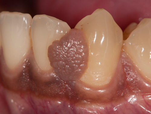
Gross Appearance of Periapical Granuloma
Granuloma represents in various forms, usually it’s a soft tissue mass that is attached to the apex of a tooth. It can be firm if the lesion is fibrous. Mass or lesion can be hemorrhagic or granular and can have dense vascular proliferation.
Clinical Presentation of Periapical Granuloma
- Sharp pain in the affected tooth if the pulp is still vital.
- If the pulp is necrotic, it might be asymptomatic with no pain on percussion
- Traumatic injury to the tooth can be seen, or deep restoration associated with pain.
- Electric pulp testing can reveal the vitality of pulp if local anesthesia is administered.
Radiographic Features seen for Periapical Granuloma
- On Periapical Radiograph, a well-defined radiolucency can usually be seen covering the root’s apical end.
- The tooth involved is clearly infected or traumatized with loss of lamina Dura at the apical end.
- In some cases, root resorption is also present
- If the tooth is root canal treated, a radiograph can appreciate the presence of any foreign particle or other factors like a failed endodontic treatment which initiated the inflammatory reaction and resulted in permanent insult to the injury.
Histopathology
- Dense Granulation Tissue containing macrophages, lymphocytes, and plasma cells.
- Chronic Inflammatory exudate (neutrophils, histiocytes, and mast cells)
- Cholesterol clefts containing multinucleated giant cells.
- Granulation tissues might be seen having scattered epithelial rest cells of Malassez.
What is the Treatment for Periapical Granuloma?
Periapical Granuloma is 100% treatable but treatment depends upon various factors like
- Cost of treatment
- Patient cooperation
- Condition of the tooth
- Patient age and history of comorbidities
If the tooth is Root Canal treated, it must be re-evaluated for a prognosis. If the prognosis is not suitable for re-treatment, extraction is the only viable option along with the lesion.
Follow-up appointments are essential in any case for a complete cure.
What is a Periapical Cyst?
Definition of Periapical Cyst
It can be defined as an inflammatory odontogenic cyst with a well-defined lining of epithelial cells derived from the rest cells of Malassez. It is also known as a Radicular cyst, dental cyst, or root-end cyst.
Pathogenesis
- Decayed/carious tooth with prolonged infection initiates
- An Inflammatory reaction, which triggers the formation of
- Rest cells of Malassez, which forms epithelium.
- Epithelium becomes necrotized
- Results in the formation of a cyst (sterile or infected)
Diagnosis of Periapical Cyst
- Periapical Radiograph or CBCT
- Clinically during dental examination
- Histopathology (whole lesion needs to be removed and examined under a microscope)
What are the Clinical findings for Periapical Cyst?
- If the cyst is sterile, it is asymptomatic.
- The tooth involved is either chronically infected or presents with failed endodontic treatment.
- Diagnosed coincidentally on a radiograph
- Swelling might be present on the outer region of the tooth if the cyst is expanding
- Pain if the cyst is infected
Radiographi Findings for Periapical Cyst
- Round or oval radiolucency on apical end on a Periapical radiograph.
- Well-defined cortical border if the cyst is not infected
- Root resorption can be seen on adjacent roots if the cyst is expanding
- Cortical border can be lost if the cyst is infected.
Histopathology
- Lining consists of stratified squamous epithelium
- Acute inflammatory exudate (neutrophils, histiocytes, mast cells, plasma cells, etc.)
- The cysts of maxillary incisors are lined by respiratory epithelium that is, the pseudo stratified ciliated columnar epithelium (due to close proximity of maxillary sinus)
- Amorphic, eosinophilic and crescent-shaped hyaline Ruston bodies can be seen
- Fibrous capsule can be appreciated
- Cyst lining contains cholesterol clefts.
What are the treatment options for Periapical Cyst?
- In small cysts (<2 cm), an infected tooth can be saved by removing the necrotic pulp along with the cyst and performing root canal treatment.
- In larger and expanded cysts, treatment options like enucleating or marsupialization are present along with tooth extraction, especially if osseous structures are involved.
Difference between Periapical Granuloma and Cyst
Definition of Periapical Granuloma vs. Cyst
Periapical Granuloma can be defined as a mass of granulation tissue or fibrous tissue present at the apical region of a tooth that is either necrosed or chronically infected, while a periapical cyst (radicular cyst) is a small oval or round-shaped sac lined by epithelial cells as a result of chronic infection of the tooth.
Pathogenesis of Periapical Granuloma vs. Cyst
Inflammatory response initiated by the necrotic pulp of the infected tooth triggers the formation of granulation tissue at the base of the root canal in the periapical granuloma. In periapical cysts, chronic decay of the tooth triggers an inflammatory reaction that further initiates the release of epithelial rests of Malassez, resulting in an epithelial lines sac called cyst. A periapical cyst is a sequalae of Periapical granuloma.
Clinical Features of Periapical Granuloma vs. Cyst
Periapical granuloma presents clinically with pulp necrosis and pain on percussion while the periapical cyst is perpetually asymptomatic and is found coincidentally on the radiograph.
Radiographic Findings of Periapical Granuloma and Cyst
Periapical radiograph shows almost similar features of both periapical granuloma and periapical cyst, however, CBCT shows slight differences in the density of borders like a sclerotic border lining the cyst but it needs to be further evaluated by histopathology and correlated clinically for a definite diagnosis.
Histopathology
Periapical granuloma shows granulation tissue that is inflamed on the microscopic examination, while cysts can be seen as a well-defined cavity lined by an epithelial wall and dense connective tissue wall.
Treatment for Periapical Granuloma vs. Cyst
Granulomas usually do not need surgical treatment and can be resolved by non-surgical root canal treatment followed by oral medications, however, cysts do not resolve on their own by medications and may require surgical removal especially if larger than 2 cm in size.
Table comparing Periapical Granuloma and Cyst
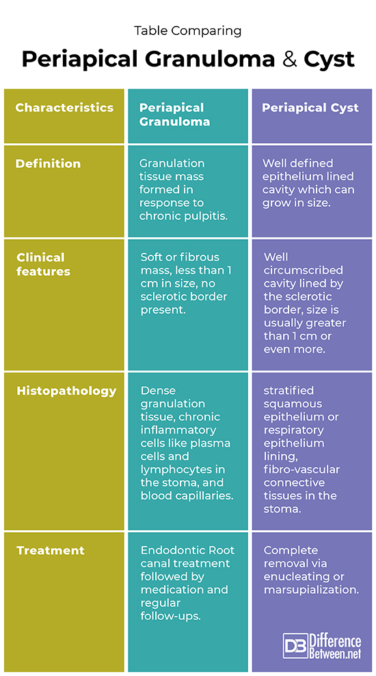
Summary of Periapical Granuloma and Cyst
- Periapical granuloma is a soft lesion present at the apical end of the root of an infected tooth while a periapical cyst is an epithelium-lined cavity.
- Granuloma is less than 1 cm in size and the periapical cyst is usually 1-2 cm and can even expand more.
- On microscopic examination, granuloma shows dense granulation tissues with predominating chronic inflammatory cells while the cyst represents a stratified squamous epithelium lining with connective tissue cells in the stoma.
- On a radiograph, they are somewhat similar in radiolucency. However, granuloma has no sclerotic border while cyst is lined by a sclerotic border.
- Treatment of granuloma is non-surgical while cyst needs surgical excavation.
FAQs
What is the difference between periapical granuloma and abscess?
Periapical granuloma is a lesion formed as a result of chronic inflammation of the decayed tooth while the abscess is the result of acute inflammation.
Which is bigger granuloma or cyst?
A cyst ranges from 1-2 cm in size and can expand even more while a granuloma is usually less than 1 cm.
Is a granuloma a cyst?
No, a granuloma is not a cyst but if it is left untreated for a prolonged period of time, can turn into a cyst.
What is a periapical granuloma?
A periapical granuloma is a small mass or lesion of granulation tissue present at the apex of a tooth infected by acute or chronic pulpitis.
How can you tell the difference between a cyst and a granuloma?
Due to similar features and radiographic appearance, the only definite way to differentiate between a cyst and a granuloma is a histopathological examination.
Should granulomas be removed?
Granulomas do not require removal; endodontic root canal treatment of the affected tooth resolves them on their own.
Should I be worried about a granuloma?
A granuloma is not as such a piece of bad news or fatal, however, treatment shouldn’t be delayed to save the tooth and bone structure.
Do granulomas keep growing?
Granulomas don’t grow beyond a certain point in size.
What is the best treatment for granuloma?
The best treatment plan is to perform Root Canal treatment of the affected tooth and regular follow-up appointments with your dentist.
- Difference Between Cystocele and Rectocele - September 8, 2023
- Comparison Between DHEA and DHEA Sulfate - September 1, 2023
- Difference Between Osteoporosis and Osteopenia - June 14, 2023
Search DifferenceBetween.net :
Leave a Response
References :
[0]Periapical (dental) granuloma. Pathology Outlines - Periapical (dental) granuloma. (n.d.). Retrieved January 16, 2023, from https://www.pathologyoutlines.com/topic/mandiblemaxilladentalgranuloma.html
[1]De Rosa, C. S., Bergamini, M. L., Palmieri, M., Sarmento, D. J. de S., de Carvalho, M. O., Ricardo, A. L. F., Hasseus, B., Jonasson, P., Braz-Silva, P. H., & Ferreira Costa, A. L. (2020, October 9). Differentiation of periapical granuloma from radicular cyst using cone beam computed tomography images texture analysis. Heliyon. Retrieved January 16, 2023, from https://www.ncbi.nlm.nih.gov/pmc/articles/PMC7560585/
[2]DE SÁ LEITE, M. A (2014). Histopathological analysis of periapical granuloma and radicular cysts: A comparative study. Oral Surgery, Oral Medicine, Oral Pathology and Oral Radiology, 117(2). https://doi.org/10.1016/j.oooo.2013.12.309