Difference Between Fistula and Shunt
What is Fistula and Shunt?
They both are designed for vascular access
Fistula
An AV fistula is surgically created by connecting an artery to a vein. It is placed in the wrist, upper arm or forearm. It is created to cause extra pressure by pushing an increase in the blood flow into the veins, making it stronger and bigger, thereby creating easy vascular access to the blood vessels.
Shunt
A shunt is a small passage created artificially and placed in the brain through surgery which allows movement of the fluid from one part of the body to another. It is made of synthetic tube tunnelled under the skin, creating a connection between the artery and the vein, and offering needle placement access for dialysis.
Difference between Fistula and Shunt
Description
Fistula
It is an abnormal connection that is created surgically creating a connection between the artery and the vein. It uses the blood cells and tissue of the body itself and no outside components. It may be surgically created to treat haemodialysis, be congenital, or acquired because of a pathologic process, like erosion of an arterial aneurysm or trauma
Shunt
Shunt is a medical device that is created artificially using outside components with no blood cells and body tissues from the body. It is usually placed in the brain or sometimes spine to drain excessive cerebrospinal fluid and divert it to another place in the body for reabsorption.
Location
Fistula
AV fistula is created and placed in your arm. In some situations, and depending upon the condition, it is even placed in the leg.
Shunt
Shunt is defined as a very tiny and hollow tube made of plastic inserted into the ventricles [cavities in the central nervous system (brain and occasionally spine), filled with fluids] on the scalp by means of incisions. The plastic tube is then passed under the skin, mostly the abdomen, to the other part of the body.
Shunt is usually inserted to help drain the excess cerebrospinal fluid and re channel it to another place in the body where it can be absorbed again. Occasionally, it can be inserted into linings of the lungs or one of the heart chambers.
Types
Fistula
The 3 most common Arteriovenous Fistulas are the;
- Brachiocephalic fistula – It is created surgically between the cephalic vein and brachial artery at the level of the cubital fossa and is linked with a 4.5-year patency rate of eighty percent.
- Radiocephalic fistula – It is created at the wrist.
- The brachial artery–to–transposed basilic vein fistula – This AV Fistula is an alternative vascular access for haemodialysis. A skin incision is made longitudinally in the interior side of the arm which is, transposed subcutaneously, connected end-to-side to the brachial artery and the basilic vein exposed.
Shunt
- Ventriculoperitoneal Shunt – drains excess cerebrospinal fluid
- Ventriculoatrial Shunt– drains excess cerebrospinal fluid into the right artrium of the heart
- Ventriculogallbladder shunt – drains excess cerebrospinal fluid into gall bladder
- Ventriculopleural shunt – drains excess cerebrospinal fluid into the chest or lungs
- Lumboperitoneal shunt – drains excess cerebrospinal fluid into the peritoneal cavity
Uses
Fistula
- In AV fistulas are created using the blood vessels and tissues from your body. There are no artificial components used.
- They have minimal possibility of becoming clotted or infected
- Easy maintenance in comparison to other access options
- They can perform and function for years.
- Arteriovenous Fistulas reduce the time required for treatment by providing good blood flow to the dialyzer.
Shunt
Useful in treatment of hydrocephalus, Idiopathic intracranial hypertension, Shunt Infection, Subdural Hematoma, Chiari malformation, hearing loss in left ear, blurry vision, haemorrhagic stroke
Complications
Fistula
Complications associated with AVFs are;
- Thrombosis – pain, tremors and absence of feeling
- Steal syndrome – pain during exercise, ischemic pain at rest, gangrene, necrosis
- Ischemic neuropathy – sensory loss, weakness in fingers and hands, paralysis of radial, ulnar and median nerves
- Aneurysm – signs of bleeding, ulceration, infection, diffuse
- Congestive heart failure (Orthopnea, Dyspnea)
- Infection – Local symptom of infection (rubor, calor, dolor)
Shunt
Complications associated with shunts are;
- Obstruction due to shunt malfunction.
- Infection, mostly in children
- Bowel perforation
- Subdural hematoma formation
- Pseudocyst formation
- Over-draining, (resulting in subdural hematoma formation)
- Multiloculated hydrocephalus
- Seizures
- Malfunctioning of shunt could cause – Vomiting, headache, tiredness, personality change, loss of coordination of balance, swelling along shunt tract
- Abdominal complications
Risk of infection
Fistula
The risk infection or clotting is less in comparison to other forms of vascular accesses.
Shunt
In case of shunt, there exists an increased risk of infections, aneurysms, and blood clotting.
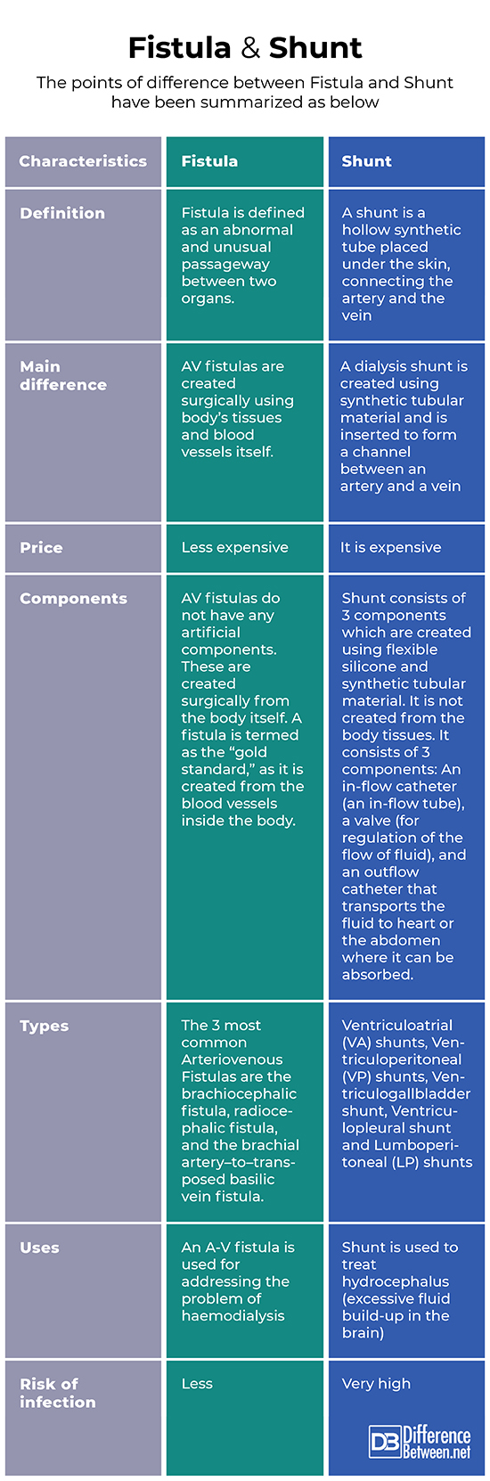
Summary
The points of difference between Fistula and Shunt have been summarized as below:
- Difference Between Global Warming and Greenhouse Effect - May 18, 2024
- Difference Between Vaccination and Immunization - March 3, 2024
- Difference Between Selective Mutism and Autism - February 25, 2024
Search DifferenceBetween.net :
Leave a Response
References :
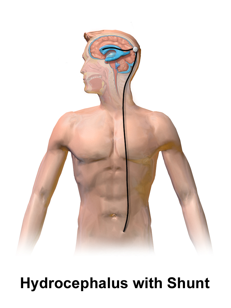
[0]Image credit: https://commons.wikimedia.org/wiki/File:Hydrocephalus_Shunt_Adult.png
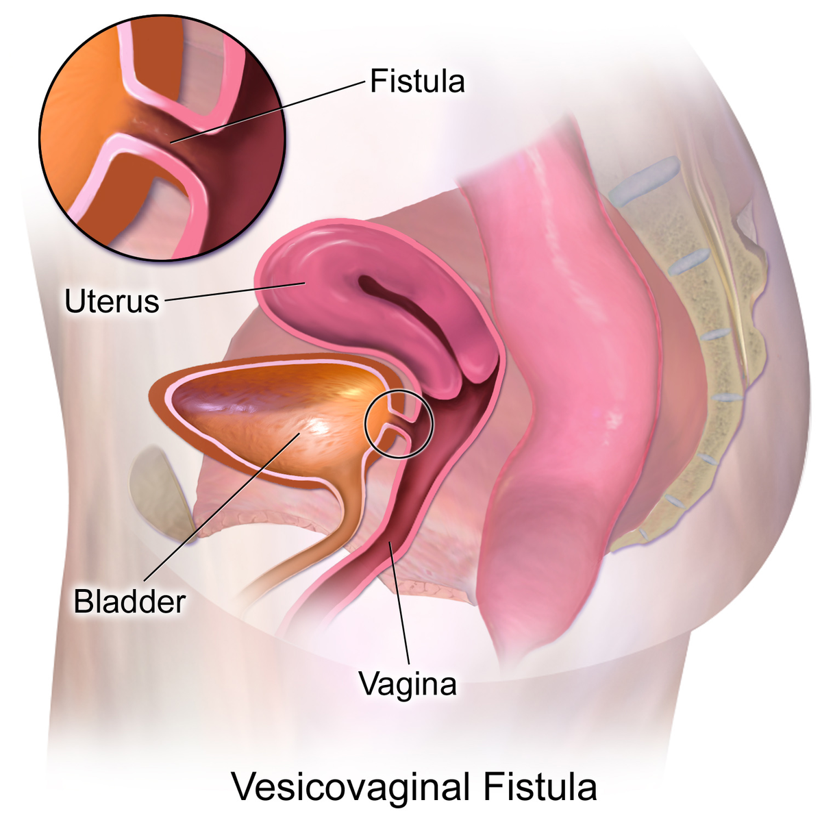
[1]Image credit: https://commons.wikimedia.org/wiki/File:Vesicovaginal_Fistula.png
[2]Al-Jaishi, A. A., Liu, A. R., Lok, C. E., Zhang, J. C., & Moist, L. M. (2017). Complications of the arteriovenous fistula: a systematic review. Journal of the American Society of Nephrology, 28(6), 1839-1850.
[3]Bansal, H., Gupta, G., Gupta, M., & Kaushal, R. (2015). Unusual ventriculoperitoneal (VP) shunt tube extrusion through anus in a child with Dandy Walker malformation: a rare case report. Journal of clinical and diagnostic research: JCDR, 9(1), PD25.
[4]Dagher, F., Gelber, R., Ramos, E., & Sadler, J. (1976). The use of basilic vein and brachial artery as an AV fistula for long term hemodialysis. Journal of Surgical Research, 20(4), 373-376.