Comparison of Pig’s Heart And Human Heart
Recently much interest has been centred towards the comparison of a pig’s heart and human heart in the pursuit of Xenotransplantation. This is because there has been a growing demand of organ transplantation which is unmet by the number of human donors (Samstein and Platt, 2001). Although, both belong to the genera of Mammalian origin there are marked difference in the anatomy of their hearts and also their associated physiological functioning. Both the hearts are divided into the auricular and ventricular chambers and have their drainage through the pulmonary arteries and aorta into the lesser and greater circulation. The myocardial volume supplied by left coronary arteries are higher and dominant compared to right coronary arteries in both these hearts as the left heart needs more oxygen supply for its greater activity to pump blood into the systemic circulation (Cooper, Gollackner and Sachs, 2002).
The coronary arteries arise from the aortic bulbus in a similar fashion in both these groups. Further the left coronary artery is very short and divides into an interventricular anterior branch and a left circumflex branch in both these species. These branches mainly give off collateral branches that supply the left atrium and the ventricles (Cooper, Gollackner and Sachs, 2002). The interventricular anterior branch gives off a proximal wide interventricular septal branch that spreads into the dorsal and central parts of the interventricular septum in both these species. The anastomoses between the various branches of coronary arteries are common to both the group of hearts (Cooper, Gollackner and Sachs, 2002). However there are quite a few differences that must be considered from a physiological and immunological standpoint before considering xenotransplantation of a pig’s heart into a human being. The comparison of both hearts is discussed as follows (Cooper, Gollackner and Sachs, 2002):
| Features | Pig’s Heart | Human Heart |
| Shape and Orientation of the heart | Possess a typical “Valentine shaped heart” which is oriented in line with the unguligrade stance of the pig | The heart is trapezoidal in shape and is oriented in line with the orthograde posture of the human being |
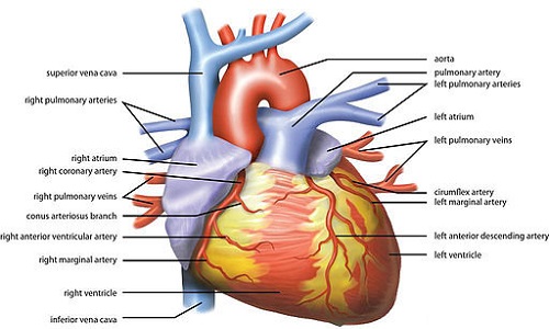
| Presence of tubular appendage | Observed in the right atrium | Observed in the left atrium |
| Orientation of the vena cava’s | The superior and inferior vena cava opens into the right atrium at right angles to each other | The superior and inferior vena cava opens into the right atrium in a straight line at 180 degree. |
| Azygous Vein | Prominent left azygous vein enters into the left side of the heart and drained via coronary sinus. | No prominent azygous vein but reduced left superior caval vein or oblique vein present |
| Characteristic of Pulmonary veins | Left atrium receives only 2 pulmonary veins | Left atrium receives only 4 pulmonary veins |
| Sweep between inlet and outlet components of right ventricle | Less prominent | More prominent |
| Muscular moderator in right ventricle | Prominent and situated higher up in the right ventricle | Less prominent and situated lower down in the right ventricle |
| Characteristic of Apical components | Contains coarse trabeculations and are broader | Coarse trabeculations are absent and is much narrower |
| Aortic-Mitral fibrous continuity | Reduced as 2/3rd of aortic valve is supported by left ventricular musculature | Not reduced |
| Length of the left and right coronary arteries | Each coronary arteries runs similar length and gives off the interventricular branch | The coronary arteries do not run similar in length hence the length and origin of the interventricular posterior branch are highly variable. |
| Coronary Dominance | Right coronary dominance absent | Surface distribution of the coronary arteries are predominantly on the right side of the heart |
| Difference in right and left atrio-ventricular branches | The right atrio-ventricular branches are less developed than their left counterparts | No such differences exist between the right and left atrio-ventricular branches |
| Recognisability of diagonal or left branches to conus arteriosus | Non recognizable | Well recognizable |
- Difference Between “Heart attack” and “Cardiac arrest” - June 24, 2016
- Difference Between Connective Tissue and Epithelial Tissue - June 22, 2016
- Difference Between Migraine And Stroke - January 11, 2016
Search DifferenceBetween.net :
1 Comment
Leave a Response
References :
[0]Cooper., D, Gollackner., B and Sachs.,D. (2002). Will the pig solve the transplantation backlog? Annu Rev Med, 53: 133-147.
[1]Samstein., B and Platt., J. (2001). Physiologic and immunologic hurdles to xenotransplantation. J Am Soc Nephrol, 12: 182-193.
[2]https://commons.wikimedia.org/wiki/File:Wiki_Heart_Antomy_Ties_van_Brussel.jpg
answered a lot of my questions